
Eaton-Peabody Laboratories
3D Virtual Model of Surgical Otologic Anatomy in Humans
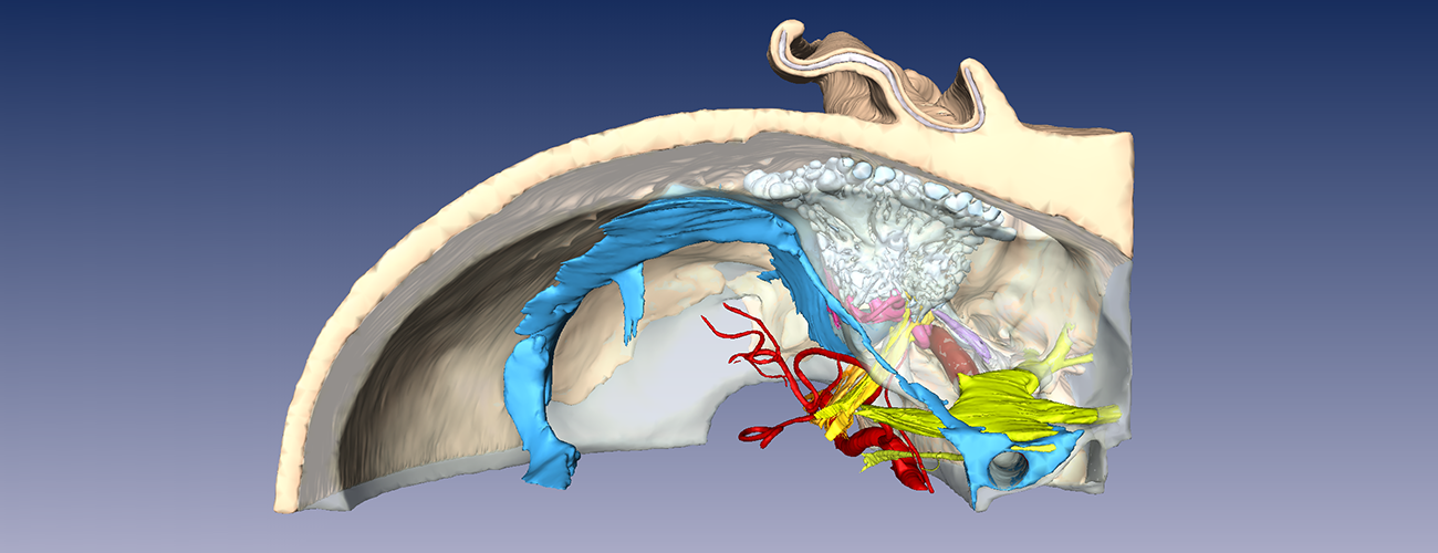
About the Visible Ear Model
The "Visible Ear" is a library of digital images of a human temporal bone and surrounding structures developed by Mads Sølvsten Sørensen, MD, and colleagues from the Department of Otolaryngology–Head and Neck Surgery, Rigshospitalet, Copenhagen, Denmark (Sorenson et al, ORL, 2002; 64:378-381).
The sections were obtained from an 85-year old female with no history of otologic disease. The temporal bone and surrounding structures were serially sectioned at 25 micron thickness. Anatomical structures of interest were segmented by Dr.Sørensen and colleagues. This data was used to generate the present model, which is a surface rendering of various structures including the external, middle, and inner ears, cranial nerves, carotid and vertebral arteries, sigmoid and cavernous venous sinuses, dura with CSF space, as well as the skin and soft tissues around the ear.
The model was generated using Amira, version 3.1. The surface model can also be sliced open in the X, Y, or Z planes and the appropriate raw histological image can be superimposed on the cleavage plane.
Installation by Operating System
- Download and uncompress the setup package
- Double click on the setup.exe file and follow the instructions on the wizard to install
- The program will automatically run after the installation
- Please refer to the user guide for how to use this software
- Download and uncompress the package
- Double click on the application to run the program
When running the program for the first time, the OS will complain that this program is from an unidentified developer. Just right click on the application, select "Open" and then choose "Open" in the pop-up window. You only need to do this the first time.
- Download and uncompress the setup package
- Double click on the setup.exe file and follow the instructions on the wizard to install
- The program will automatically run after the installation
Contributors to this model include Dr. Sørensen, Haobing Wang of Mass Eye and Ear, and Michael Teixido, MD, and Brian Kung, MD, of Delaware Biotechnology Institute. Model development was supported by a core grant from the NIDCD (P30 DC05209). Michael Teixido, MD, has additionally created a set of teaching movies based on the 3D virtual models of the human temporal bone.