
Eaton-Peabody Laboratories
3D Virtual Model of a Human Temporal Bone
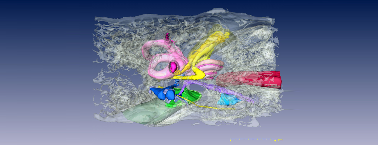
About the Temporal Bone Model
This 3D virtual model of a human temporal bone is a powerful teaching tool for learning the complex anatomy of the human temporal bone and for relating the 2D morphology from a histological section to the 3D anatomy.
The model was created from archival histological sections from a 14-year-old male. The specimen was formalin fixed, decalcified, embedded in celloidin, serially sectioned in the axial plane at 20 microns, stained with hematoxylin and eosin, and mounted on slides. Low-power views of every fifth section through the temporal bone were digitized and imported into Amira v3.1 (Mercury Computer Systems/TGS, San Diego, CA). The sections were aligned and segmented into anatomical structures of interest.
The model is a surface rendering of the following structures of interest, which currently includes (among others):
- Bone and air spaces of the temporal bone
- Perilymphatic and endolymphatic spaces (including the cochlear aqueduct and endolymphatic duct and sac)
- The sensory epithelia of the cochlear and vestibular labyrinths
- The ossicles and tympanic membrane
- Middle-ear muscles
- The carotid artery
- Auditory, vestibular and facial nerves
For each of these structures, the surface transparency can be individually controlled, thereby revealing the 3D relations between surface landmarks and underlying structure. New structures of interest can be added within the Amira software.
The 3D surface model can also be sliced open at any section and the appropriate raw histologic image superimposed on the cleavage plane. Leafing through the section stack in this way provides a powerful view of the relation between microscopic images and 3D anatomy. The image stack can also be re-sectioned in any arbitrary plane.
Installation by Operating System
- Download and uncompress the setup package
- Double click on the setup.exe file and follow the instructions on the wizard to install
- The program will automatically run after the installation
- Please refer to the user guide for how to use this software
- Download and uncompress the package
- Double click on the application to run the program
When running the program for the first time, the OS will complain that this program is from an unidentified developer. Just right click on the application, select "Open" and then choose "Open" in the pop-up window. You only need to do this the first time.
- Download and uncompress the setup package
- Double click on the setup.exe file and follow the instructions on the wizard to install
- The program will automatically run after the installation
The model was made under the supervision of M. Charles Liberman, PhD, and Saumil Merchant, MD, by Haobing Wang and Clarinda Northrop of Mass Eye and Ear. Model development was supported by a core grant from the NIDCD (P30 DC05209).