
Eaton-Peabody Laboratories
Imaging Core
The human temporal bone contains a large number of complex structures within a small space. This complex anatomy is a challenge for students in the basic science and medical disciplines. Computer-based 3D virtual models have the potential to become valuable tools for teaching and learning this anatomy.
The Imaging Core of the Eaton-Peabody Laboratories at Mass. Eye and Ear has developed several such models and made them available as downloadable freeware for teaching and educational purposes.
Downloadable 3D Virtual Models
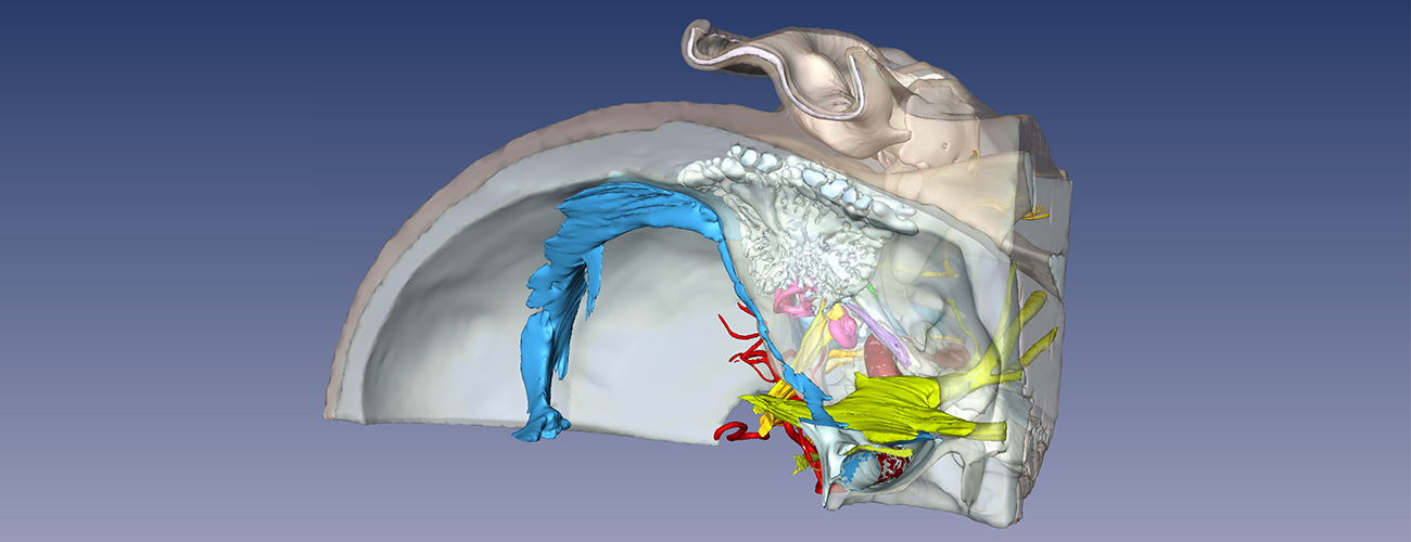
- The Visible Ear: Developed from a library of visual images from an 85-year old female, this model contains the entire human temporal bone and surrounding structures of the skull base.
- Temporal Bone Model: Developed from archival histologic sections of a 14-year old male, this model depicts the majority of the structures of the external, middle, and inner ears, along with the mastoid air spaces.
- Round Window Model: A more detailed subset of the Temporal Bone Model. This depicts the detailed anatomy of the round window membrane and adjacent structures of the cochlea.
- Incudostapedial Joint Model: Another more detailed subset of the Temporal Bone Model, which depicts the anatomy of the distal incus, incudostapedial joint, and head of stapes.
These models were supported by grants from the National Institutes on Deafness and Communication Disorders (NIDCD) and the NIDCD National Temporal Bone Registry. The Visible Ear was developed by Mads Sølvsten Sørensen, MD, and colleagues from the Department of Otolaryngology–Head and Neck Surgery at Rigshospitalet, University of Copenhagen, Denmark.